| Proteins
that make up the human body are widely diverse and their
structure is closely related to how the protein works
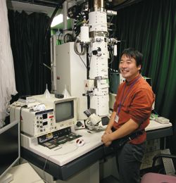
and so how the body functions. The electron microscope
is a powerful tool for elucidating the relationship
between protein structure and its function. It is now
possible to analyze photographic data from electron
microscopes to determine the 3D structure of proteins
including their own structure and complexes. This macromolecule
analysis technique can be also used for the development
of pharmaceuticals and nano-products. In this program,
students are given the opportunity to actually use one
of only two high-performance electron microscopes in
the world and to learn the basic skills needed to operate
it. They will also photograph protein complexes and
reconstruct 3D images by digital image processing. The
constructive proposals from students are welcome to
examine other things than proteins under the microscope.
This is one of the most appropriate program for students
who want to observe something under an electron microscope.
|
 |
 |
|